
IMAGING IN INJURY ASSESSMENT
The topic of imaging in musculoskeletal medicine is vast and difficult to cover in one article. It is however important to discuss the various reasons for different types of imaging and where they fit in when trying to figure out what is happening with your injury. Everyone often assumes an MRI is the best form of imaging available. Whilst at times this may be appropriate often there may be another test which is more suitable for a particular condition. Cost of imaging, medicare referring rights, and patient suitability all play a role in deciding what imaging to send you for.
Some principles around if imaging should be used in your case include:
The investigation should change or influence your management.
Is the test worth getting and will it change your management? The basis for treating any musculoskeletal condition is working off an accurate diagnosis. Any investigation incurs not only cost (to you and Medicare), but also a time cost in going and getting it done, so it needs to be helpful to your particular management. There is also some imaging that can open create more issues, such as feeding chronic pain thoughts and potentially prolonging symptoms.
The investigation should be the most appropriate one to diagnose the issue based on YOUR presentation
There should be a consideration of all types of imaging relevant to the patient before a modality is chosen. This will be driven your history, but more importantly your clinical examination. The test should always try to confirm the clinical suspicion, not make the diagnosis on its own.
What type of imaging is there?
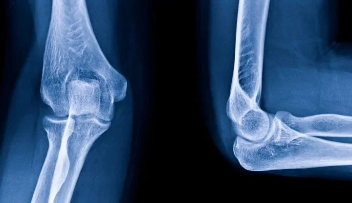
X-ray: Often, the first type of imaging used is a plain X-ray. An X-ray will convey important information with respect to bone and joint issues, particularly osteoarthritis, and assess for unexpected more serious pathologies, such as a cancer. Stress or weight-bearing views may also provide important information in joint instability, such as in a Lis Franc injury in the foot. It is also the cheapest type of imaging, with many sequences significantly covered by Medicare. However, if purely assessing soft tissue structures, such as muscle or ligament, there are more suitable imaging modalities that should be used instead.
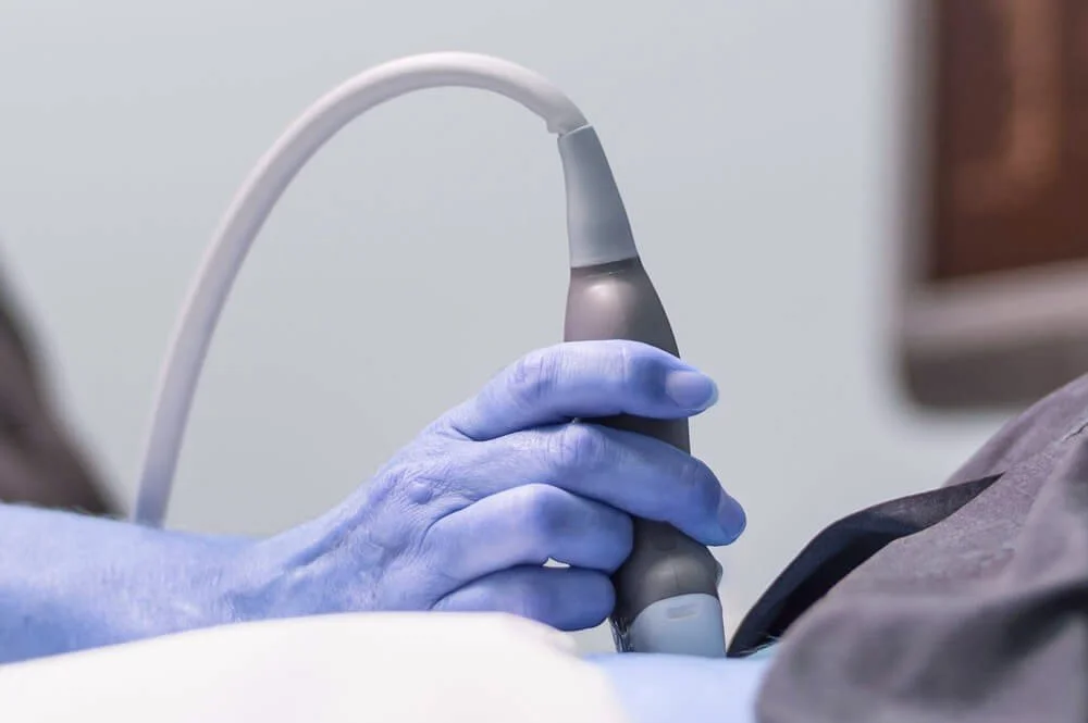
Ultrasound: Often, unfortunately, ultrasound is an overutilized modality. Whilst it can provide information in certain conditions, a proportion of these can be diagnosed purely on clinical grounds, eg. tennis elbow or plantar fascitis. The difficulty with ultrasound is it is often hard to differentiate between tears, tendinopathy etc as both pathologies reflect the soundwaves in a similar manner and therefore look the same. Ultrasound lacks the accuracy of MRI, especially with deeper tissue structures, and can be heavily dependent on the operator and the quality of the machine. One advantage of ultrasound however, can be the dynamic nature of the test, allowing assessment of the abnormality with motion in real time.
CT scan: CT scanning is most useful for assessment of bony structures and plays little role for soft tissue injury. It can be helpful for diagnosis of missed or difficult to identify fractures as an extension of plain X-ray when there is still some clinical suspicion of a fracture. A downside of CT scanning is the radiation dose, which is significantly higher than other forms of imaging, especially if performed axially.
Nuclear medicine (Bone scan): Whilst nuclear medicine imaging has been replaced in many areas by MRI, it still has a role to play in certain circumstances. Its main advantage is sensitivity, but not necessarily specificity. This means it I helpful in ruling out conditions, such as stress fractures, but MRI is generally as sensitive for this purpose. With multiple joint issues bone scanning also has the ability to look at the ‘whole body‘ at the one time, with one scan.
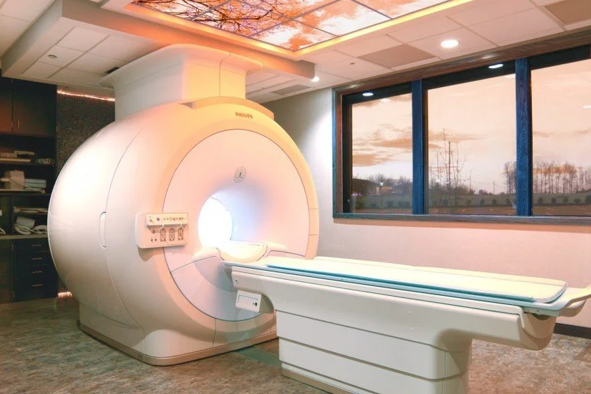
An MRI machine
MRI: MRI is clearly the pinnacle of imaging. In many ways this can be to its detriment, because it shows everything, including normal age related changes such as disc changes etc, so your clinician needs to be correlate findings with your clinical findings. As such the interpretation of the MRI is just as important as the scan report itself. MR’s strength is with soft tissue injury and its main weakness is with bony injury and particularly fractures, where CT is superior. It is, however, quite sensitive in detecting bone marrow oedema or ‘bone-bruising’.
The major downside with MRI scanning is cost. An MRI is many times more expensive than a plain X-ray and some justification needs to be made for its use from a health economic point of view. It is most beneficial when other modalities have failed to provide an accurate diagnosis or there is ‘diagnostic doubt’.
If you need help to diagnose your injury or pain, head online to book in today. Portside Sports Physio’s team will take the time to assess you and what your injury specifically needs to return you to what you love.